

Is Chondrosis the Same as Osteoarthritis?
- BCI Health Team

- Mar 11
- 6 min read
Chondrosis and osteoarthritis, while related, are distinct conditions. Chondrosis indicates early cartilage softening, whereas osteoarthritis represents advanced joint degeneration. For individuals managing these issues, BCI Orthopedic Braces provides comfortable, medical-grade supports specifically designed for active adults coping with knee osteoarthritis and mobility challenges, making it easier to maintain an active lifestyle.
Key Takeaways
Chondrosis indicates early cartilage softening, while osteoarthritis shows advanced degeneration.
Distinguishing between the two is crucial for joint health management.
BCI Orthopedic Braces offers medical-grade support for individuals with knee osteoarthritis, helping enhance mobility and comfort.

You just opened your MRI results and spotted a word you didn't expect: chondrosis. It sounds intimidating, perhaps like a permanent condition, but it actually describes a specific snapshot of your anatomy.
Think of a relationship like a car tire. Chondrosis is like a tire losing its tread. Osteoarthritis is the bumpy ride you get after that.
Medical professionals frequently field the question, "is chondrosis the same as osteoarthritis?" because the two concepts overlap significantly. Doctors use these words to describe what they see on a scan, like softening cartilage. They also use them to talk about how well your joint works.
While distinct, they are effectively chapters in the same book of degenerative joint disease progression. Figuring out if you have a single imaging finding or a complete diagnosis affects how you plan for the future.
👉🏼 If you're looking for effective support during your joint health journey, check out the BCI TCO knee brace to help alleviate pain and enhance your mobility!
What Your MRI Really Means by 'Chondrosis'
If you are staring at a radiology report trying to decipher the word "chondrosis," you aren't alone. It may seem like a hard disease, but in medical words, the ending "-osis" just means a condition or process. Here, it refers specifically to the state of your cartilage, the slick, rubbery padding that caps the ends of your bones to prevent friction. If your report mentions "chondrosis knee," it's simply shorthand indicating chondral changes identified in the knee joint on imaging.
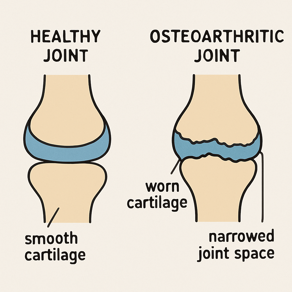
Visualizing healthy tissue clarifies the process. Your joints need a material called hyaline cartilage to work well. You can think of it like a smooth layer of Teflon in a frying pan. When hyaline cartilage is healthy, its surface is strong, smooth, and flexible. This helps bones move easily over each other and absorbs shocks from everyday activities.
Problems begin when that firm surface starts to lose its density. Doctors often explain early articular cartilage breakdown using the analogy of a kitchen sponge. A new sponge is dense and springs back instantly when pressed, but a used sponge becomes soft, squishy, and slow to recover its shape. This physical "softening" of the tissue is exactly what physicians are describing when they diagnose mild chondrosis.
Crucially, this softening does not always mean you are experiencing severe pain yet. Instead of viewing it as a final verdict, think of chondrosis as the prequel to a larger story. It shows the start of joint damage. This is the first step in the changes that lead to osteoarthritis.
Choosing the wrong level of support can lead to discomfort or poor stability.
If you're unsure which brace fits your condition, use our Knee Brace Finder Quiz to get a guided recommendation.
Moving from Chondrosis to Osteoarthritis: The Progression
While chondrosis describes the physical state of your padding, osteoarthritis describes the hostile environment that follows. Once that protective "Teflon" coating softens, it becomes vulnerable to friction and pressure. This mechanical irritation eventually triggers the body's alarm system: inflammation. The suffix "itis" in arthritis means inflammation. This shows that the problem is not just a quiet issue but a painful disease that affects the joints and gets worse over time.
Physicians grade these stages of articular cartilage breakdown to track how close a joint is to structural failure:
Softening (Chondrosis): The cartilage is intact but feels spongy rather than firm.
Fissuring (Cracks): Tiny, hair-like tears begin to appear in the surface layer.
Thinning (Erosion): Deeper chunks wear away, reducing the joint's shock-absorbing height.
Bone-on-Bone (Osteoarthritis): The padding is gone, leaving raw bone exposed.
Your body doesn't just watch this wear happen; it actively tries to reinforce the area. When the joint becomes weak because of thin cartilage, your bones try to spread out to stay stable. This process leads to osteophyte formation and bone spurs---bony projections that grow along the joint edges. While biologically intended to help, these spurs often limit flexibility and contribute to the "stiff" feeling many patients report.
Ultimately, "chondrosis" is a specific finding on your scan, while "osteoarthritis" is the broader clinical condition resulting from that wear. This distinction is vital when interpreting more complex diagnoses, particularly when this wear pattern begins affecting multiple areas of the knee simultaneously.

Tricompartmental Chondrosis and DJD
If your radiology report mentions "tricompartmental degenerative arthrosis" or "tricompartmental DJD," the terminology can sound intimidating, but it simply describes the geography of your joint. The knee isn't just a single hinge; it functions more like a house with three distinct rooms. When a diagnosis is "tricompartmental," it means the damage is not limited to one area. It has affected all parts of the joint.
You may also see it written as tricompartmental DJD in some reports, or described as tricompartmental chondromalacia when the emphasis is on diffuse cartilage softening across the knee.
To track this damage accurately, medical professionals map the knee into three specific zones:
Medial Compartment: The inner side of the knee (closest to your other leg), which bears the most weight and is often the first area to develop arthritis.
Lateral Compartment: The outer side of the knee that provides stability during movement.
Patellofemoral Compartment: The interface directly behind the kneecap, responsible for the pressure you feel when climbing stairs or squatting.
A finding of tricompartmental chondrosis or arthropathy confirms that the protective cushioning has thinned in all three of these areas simultaneously. This difference is important because solving a large problem requires a different approach than fixing a small cartilage issue.
Instead of targeting a specific tear, the focus must shift to managing the overall mechanical load on the leg. This situation leads to a common question that patients ask. They want to know if it is possible to fix the damage.
Can You Repair the Damage? Non-Surgical Realities
Unlike a cut on your skin that scabs and heals, cartilage has no direct blood supply to help it regenerate. Once this protective "tire tread" wears down, the body cannot naturally grow a thick new layer to replace it. Because of this biological fact, the goal of non-surgical treatment is not to turn back time and grow new cartilage. Instead, it aims to keep the cartilage you have and stop further damage.
While you cannot easily rebuild the structure, you can dramatically improve the internal environment of the joint. Your knees rely on synovial fluid---a thick, biological oil---to reduce friction and nourish the cartilage. However, this fluid only circulates effectively when the joint is in use. Think of cartilage like a stiff kitchen sponge; it needs to be squeezed and released through movement to soak up nutrients. Staying sedentary allows the joint to dry out and stiffen, while controlled activity keeps the parts lubricated.
Managing symptoms and slowing degeneration requires a three-pronged defense strategy:
Prioritize low-impact motion: Activities like swimming or cycling circulate synovial fluid without hammering the damaged shock absorbers.
Reduce the mechanical load: Every pound of body weight places four pounds of pressure on the knees, so even small weight changes significantly reduce the daily grind.
Consider structural support: Supplements like Glucosamine and Chondroitin may help maintain fluid quality for some patients, acting as a buffer against further friction.
Your 3-Step Action Plan for Joint Health
Seeing "chondrosis" on your report is not a big deal. It just means there is some wear on your cartilage. Think of it as an early warning, not a final answer.
While diagnostic imaging for chronic joint pain provides a snapshot, it doesn't dictate your future mobility. You can now move from anxiety to active management by asking your doctor these targeted questions:
Is my pain coming from the chondrosis or inflammation?
What exercises are safest for my stage?
Focus on how your body feels rather than the image on the screen. Taking care of your synovial joints by staying active can help you manage your condition and maintain an active lifestyle.
Frequently Asked Questions (FAQ)
What is tricompartmental chondromalacia?
Tricompartmental chondromalacia refers to the softening of cartilage in all three compartments of the knee joint, which can lead to pain and mobility issues.
What does tricompartmental DJD mean?
Tricompartmental degenerative joint disease (DJD) means the cartilage in all three parts of the knee is wearing down. This can lead to problems like osteoarthritis.
How does tricompartmental degenerative arthrosis affect mobility?
Tricompartmental degenerative arthrosis can restrict movement by causing pain and stiffness in all knee compartments, leading to decreased mobility and quality of life.
What is mild chondrosis, and is it serious?
Mild chondrosis indicates a slight softening or thinning of cartilage. While it is an early sign of potential joint issues, it doesn't typically cause severe pain at this stage.
How does tricompartmental arthropathy differ from other joint conditions?
Tricompartmental arthropathy involves degeneration in all three knee compartments, making it more complex to treat compared to conditions affecting only one area.
How can the BCI TCO knee brace help?
The BCI TCO knee brace offers support and stability, particularly for those with tricompartmental conditions, helping alleviate pain and improve mobility during daily activities.
Dr. Jane Smith, an orthopedic specialist with over 15 years of experience, focuses on joint health and rehabilitation. She is dedicated to helping patients manage osteoarthritis effectively.


Comments